Dentists use radiographs for many reasons. In the Bisecting Angle Technique the x-ray beam is directed perpendicular T shape to an imaginary line which bisects divides in half the angle formed by the long axis of the tooth and the long axis of the film.
Periapical Radiography Clinical Gate
Dental x-ray unit select one a.
. The global Dental Imaging market size is estimated to be worth US 24251 million in 2022 and is forecast to a readjusted size of US 38179 million by 2028 with a CAGR of 79 from 2022-22. A dental x-ray unit permits. XLH is the most.
Floor mounted dental radiograph generator c. Digital based system Direct digital sensor or Phosphor plate Desktop or laptop computer Dental x-ray units are usually mounted on a wall overlooking the dental suite. Dental radiographs are commonly called X-rays.
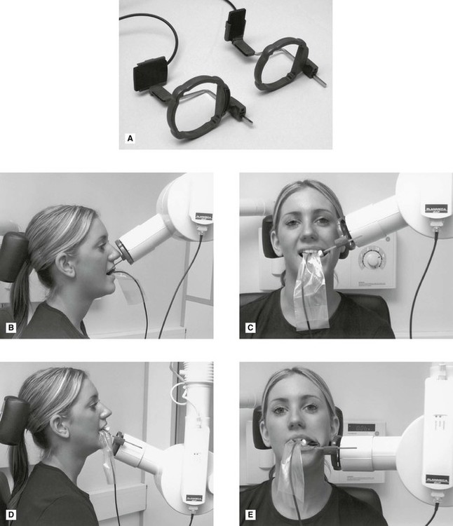
The X-ray machine is positioned alongside your head to record images of your mouth. Some dental practices have a separate room for X-rays while others perform them in the same room as cleanings. The snap-a-ray is used.
A radiographic image is formed by a controlled burst of X-ray radiation which penetrates oral structures at different levels depending on varying anatomical densities before striking the. Faulty radiographs melbia shine. X-linked hypophosphataemia XLH is an X-linked dominant disorder caused by mutations in PHEX located at Xp221 which encodes a cell-surface-bound protein-cleavage enzyme phosphate-regulating neutral endopeptidase PHEX predominantly expressed in osteoblasts osteocytes and teeth odontoblasts and cementoblasts.
Using the plaster models of 441 cases they measured and recorded the widths of all the mandibular teeth including the first. Presentation of diagnosis related to cancer by heena goverment nursing college. Ballard and Wylie were so concerned about the distortions of the X- ray films that they devised a scheme for estimating the widths of the mandibular canine and the premolars on the basis of the combined widths of the four lower incisors.
Artifact and errors in intraoral periapical radiographppt jyoti sharma. When comparing the two periapical techniques the. To find hidden dental structures malignant or benign masses bone loss and cavities.
Size 2 Film is used for Anterior and Posterior X-rays when Bisecting. X ray films hmdali. They can also be free standing or mobile and a handheld unit is now available.

Periapical Radiography Pocket Dentistry

How Make Periapical X Ray

Lecture 6 Paralleling Technique Intro Dental Radiography I
Periapical Radiography Clinical Gate

How Make Periapical X Ray

Periapical Radiography Pocket Dentistry
Periapical Radiography Pocket Dentistry
Periapical Radiography Clinical Gate
0 comments
Post a Comment